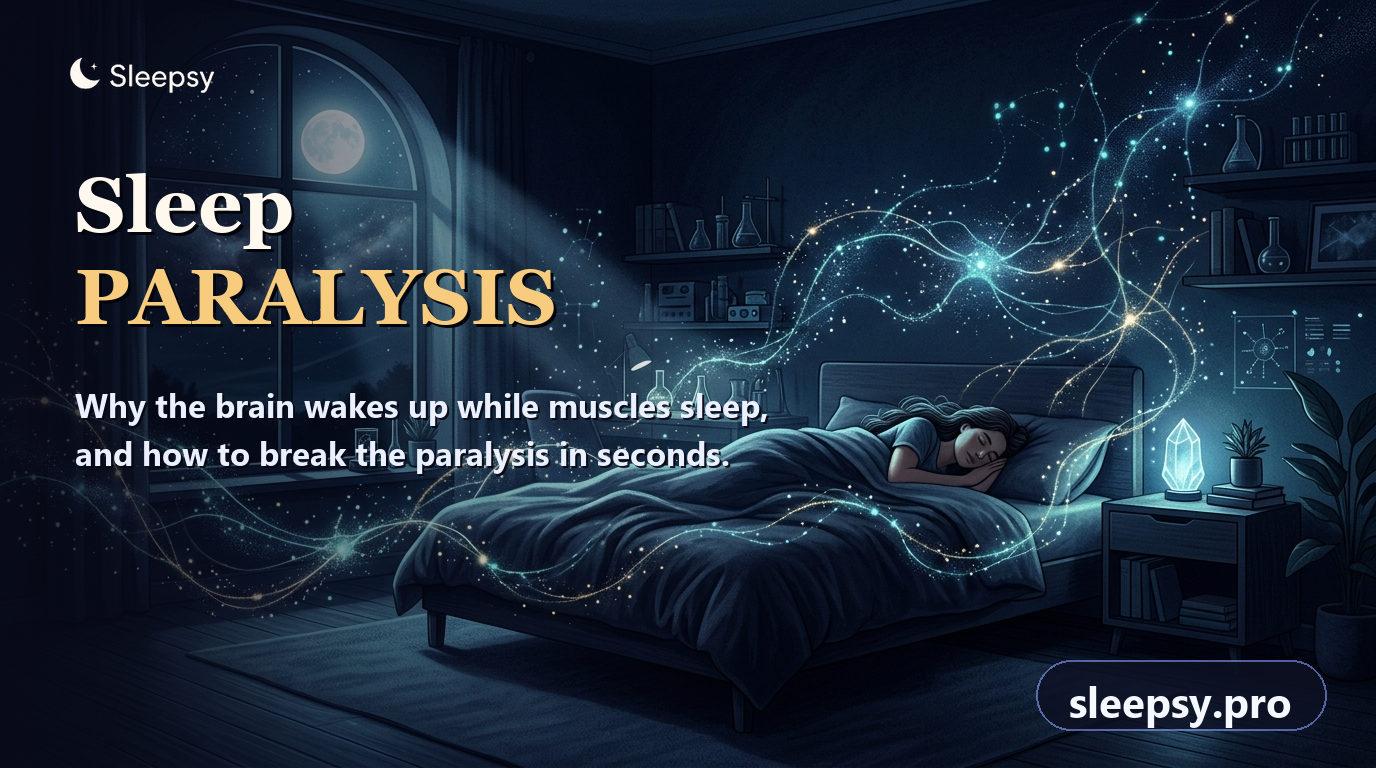
You open your eyes into the stillness of your bedroom, take in the familiar contours of the room, and suddenly realize with surging dread that your body will not move. You cannot lift an arm, turn your head, or utter a sound, while a sensation of suffocating weight settles across your chest. In the dim twilight, a sinister shadow seems to linger just at the edge of your peripheral vision.
Millions of people encounter this jarring phenomenon at least once in their lives. For centuries, folklore attributed it to nocturnal demons, witches, or supernatural visitations. Modern sleep medicine reveals a much simpler reality: sleep paralysis stems from a temporary miscommunication between a waking cerebral cortex and the spinal motor switches that guard rapid eye movement (REM) sleep.
In clinical sleep science, this episode is recognized as a harmless timing glitch. Understanding the neurochemistry behind this transition removes the dread, disarms the illusion of presence, and gives you actionable tools to regain full mobility in seconds.
Anatomy of the Glitch: What Happens in the First Seconds
In somnology, this state is termed isolated sleep paralysis. It occurs most frequently during hypnopompic transitions—the threshold where the brain awakens from REM sleep before the spinal cord has turned skeletal muscle tone back on.
In a comprehensive systematic review led by Brian Sharpless, lifetime prevalence of isolated sleep paralysis was determined to be approximately 7.6% in the general population, rising to 28.3% among university students: Sharpless & Barber, 2011.
This creates a sharp sensory-motor mismatch. Your visual cortex registers your actual bedroom walls and your auditory system hears passing street sounds, but outgoing motor commands dissolve before reaching your limbs. You feel trapped within an unresponsive physical shell.
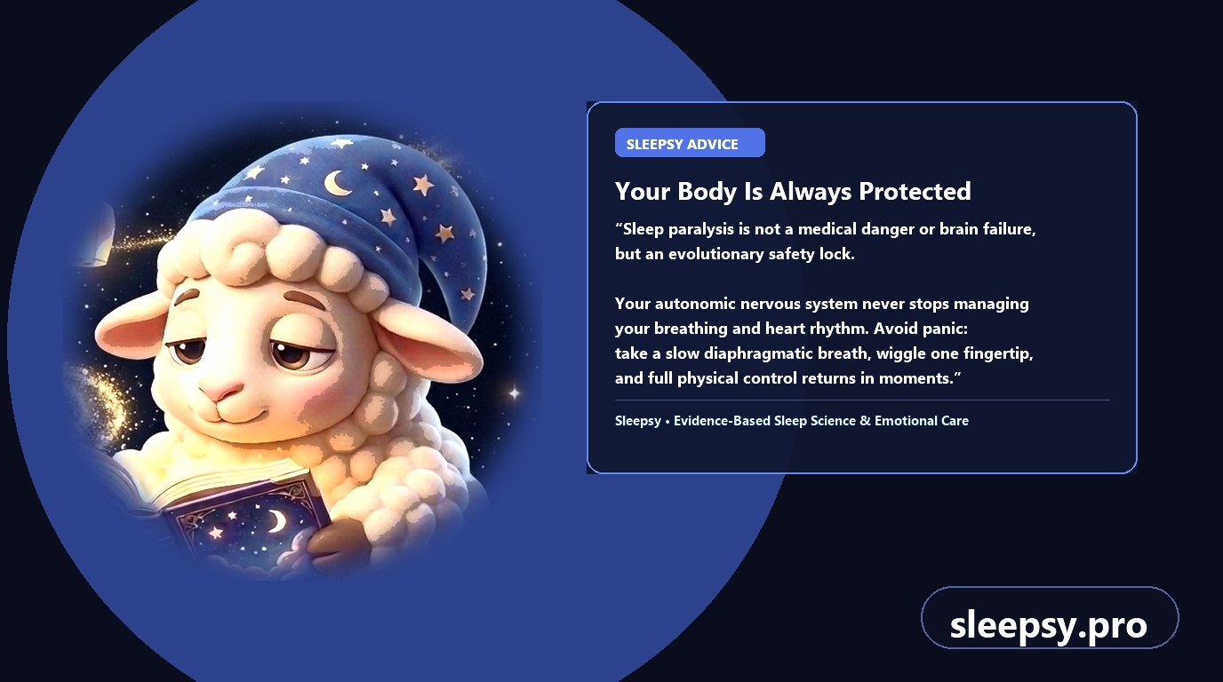
An episode typically lasts between 15 seconds and two minutes. It poses zero danger to cardiovascular or pulmonary function: autonomic pathways continue to regulate cardiac output and diaphragmatic breathing without interruption.
The Neurobiology: How GABA and Glycine Lock Skeletal Muscles
Every night, our brain cycles through non-REM and REM stages. Vivid dream narratives unfold primarily during REM sleep, a state marked by intense cerebral activity. To explore this architecture further, see our analysis of sleep cycles and dream recall.
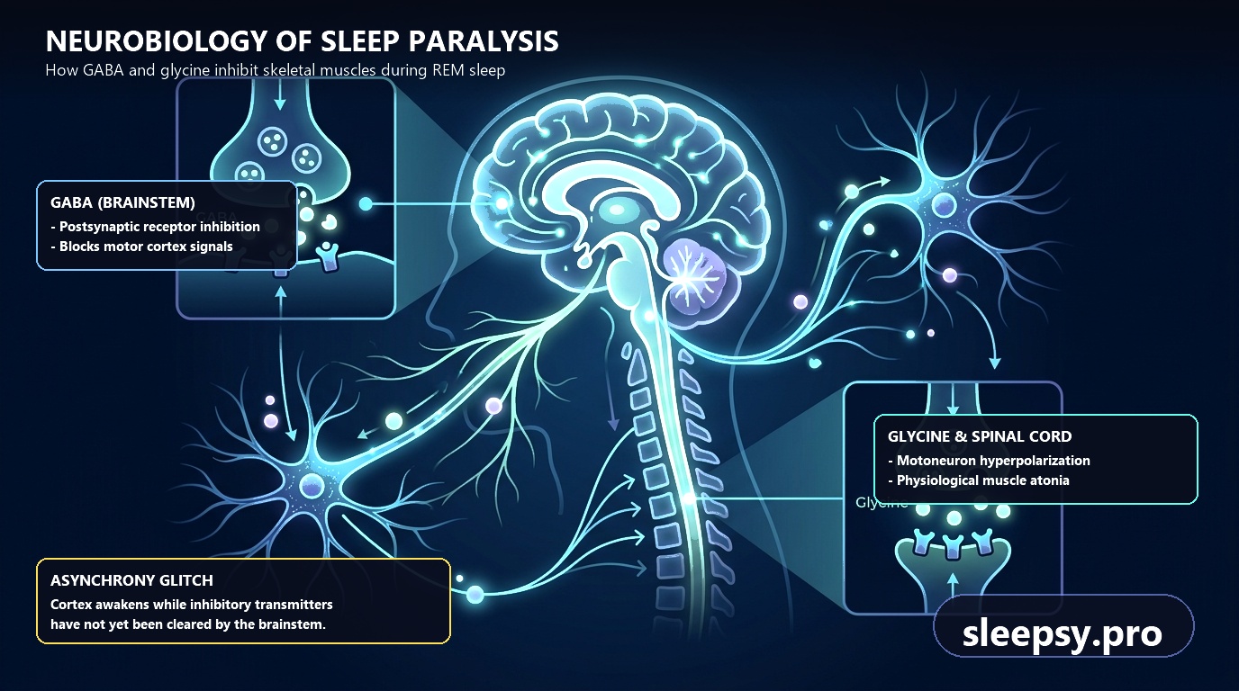
During REM dreaming, the motor cortex fires with identical intensity to waking movement. If our limbs mirrored every dream impulse, we would physically act out nocturnal events and risk severe physical injury. To prevent this, brainstem nuclei enforce a profound state of flaccid muscle inhibition known as REM atonia.
Researchers led by Olivier Clément and François Brischoux in Nature Communications demonstrated that skeletal motor neuron inhibition during REM sleep is driven by GABAergic and glycinergic medullary neurons that suppress muscle tone: Brischoux et al., 2018.
These medullary projections flood spinal motor neurons with inhibitory neurotransmitters, dropping muscle tone to zero. In sleep paralysis, a micro-asynchrony occurs: conscious awareness reboots immediately, while the biochemical clearance of GABA and glycine lags behind by several dozen seconds.
Sensory Conflict: Why the Brain Projects Shadow Figures and Chest Pressure
Physical immobility is disorienting on its own, but the acute terror that accompanies sleep paralysis is what makes it so unforgettable. Why does an immobilized mind conjure menacing apparitions in a familiar room?
The primary driver is acute hypervigilance in the amygdala—the brain’s threat detection center. Conscious awareness recognizes that voluntary muscles are unresponsive. The amygdala interprets this sudden lack of motor feedback as an existential emergency and initiates an adrenaline surge. Under ordinary circumstances, fear triggers a fight-or-flight response; here, physical movement remains blocked.
Respiratory biomechanics amplify this alarm. In REM sleep, intercostal rib muscles are inhibited, leaving ventilation entirely to the autonomic diaphragm. Attempting a voluntary, deep chest gasp meets resistance from the resting chest wall. The brain interprets this resistance as external suffocation, synthesizing the historic illusion of a crushing weight or dark figure on the chest.
Scrambling to rationalize why adrenaline is surging without an identifiable waking hazard, the hyperactive cortex weaves residual REM imagery into the bedroom setting. The result is a waking dream projection—a shadowy silhouette, auditory footsteps, or tactile vibrations.
Core Triggers: What Disrupts the REM Switch
Isolated sleep paralysis is not an illness or structural disorder; it is a physiological response to circadian misalignment and autonomic overload. Several consistent lifestyle variables lower the threshold for these transition glitches.
A systematic review of predisposing risk factors by Dan Denis and colleagues confirmed that sleeping in the supine position, sleep fragmentation, and elevated anxiety significantly increase the frequency of sleep paralysis episodes: Denis et al., 2018.
| Trigger Variable | Physiological Pathway | Corrective Strategy |
|---|---|---|
| Supine Sleeping Posture | Lying flat on the back causes the tongue and soft palate to shift rearward, compromising upper airway flow and causing abrupt micro-arousals directly out of REM sleep. | Practice side-sleeping (lateral posture); utilize an ergonomic contour pillow to support cervical alignment. |
| Chronic Sleep Deprivation | Cumulative rest deficits trigger a rebound effect, compressing REM cycles and increasing the frequency of unstable sleep-wake transitions. | Maintain a steady sleep window of 7 to 8 hours nightly to normalize REM phase density. |
| Circadian Irregularity | Rotating night shifts, variable sleep schedules, and travel across time zones desynchronize core body temperature from melatonin release. | Anchor your wake-up time to a consistent 30-minute window every day of the week. |
| Autonomic Stress Load | Elevated evening cortisol maintains cortical arousal networks while brainstem sleep centers remain locked in atonia. For deeper context, read about anxiety dreams and stress. | Establish a 45-minute digital wind-down buffer before bed to down-regulate sympathetic activation. |
Clinical Scenario: Shift Work and Nocturnal Anticipation
Consider the typical clinical pattern observed in healthcare workers or rotating shift professionals. Frequent overnight schedules, fragmented nap windows, and high cognitive responsibility steadily erode circadian stability.
In one illustrative scenario, a pediatric emergency resident began experiencing weekly episodes after transitioning to rotating night shifts. Collapsing onto an on-call cot on her back, she would awaken to discover her limbs frozen and perceive an ominous figure standing in the doorway. Although her medical training explained the physiological atonia, the sheer autonomic surge produced anticipatory bedtime phobia, leading her to sleep with overhead lights on and deepening her exhaustion.
By transitioning strictly to lateral side-sleeping, stabilizing baseline sleep opportunity, and applying peripheral micromovement drills, the episodes resolved. Understanding that the perceived apparition was merely an amygdala misattribution neutralized the fear cycle.
The 4-Step Protocol: How to Break Sleep Paralysis in Under 30 Seconds
The most common instinct during sleep paralysis is to fight back—straining with maximum force to throw yourself out of bed or scream. Because spinal pathways are chemically gated, this struggle only feeds the panic loop and prolongs the episode.
In an interventional clinical cohort study, Baland Jalal and co-authors demonstrated the efficacy of emotional decoupling and muscle micro-relaxation (Meditation-Relaxation therapy) in rapidly breaking sleep paralysis episodes: Jalal et al., 2020.
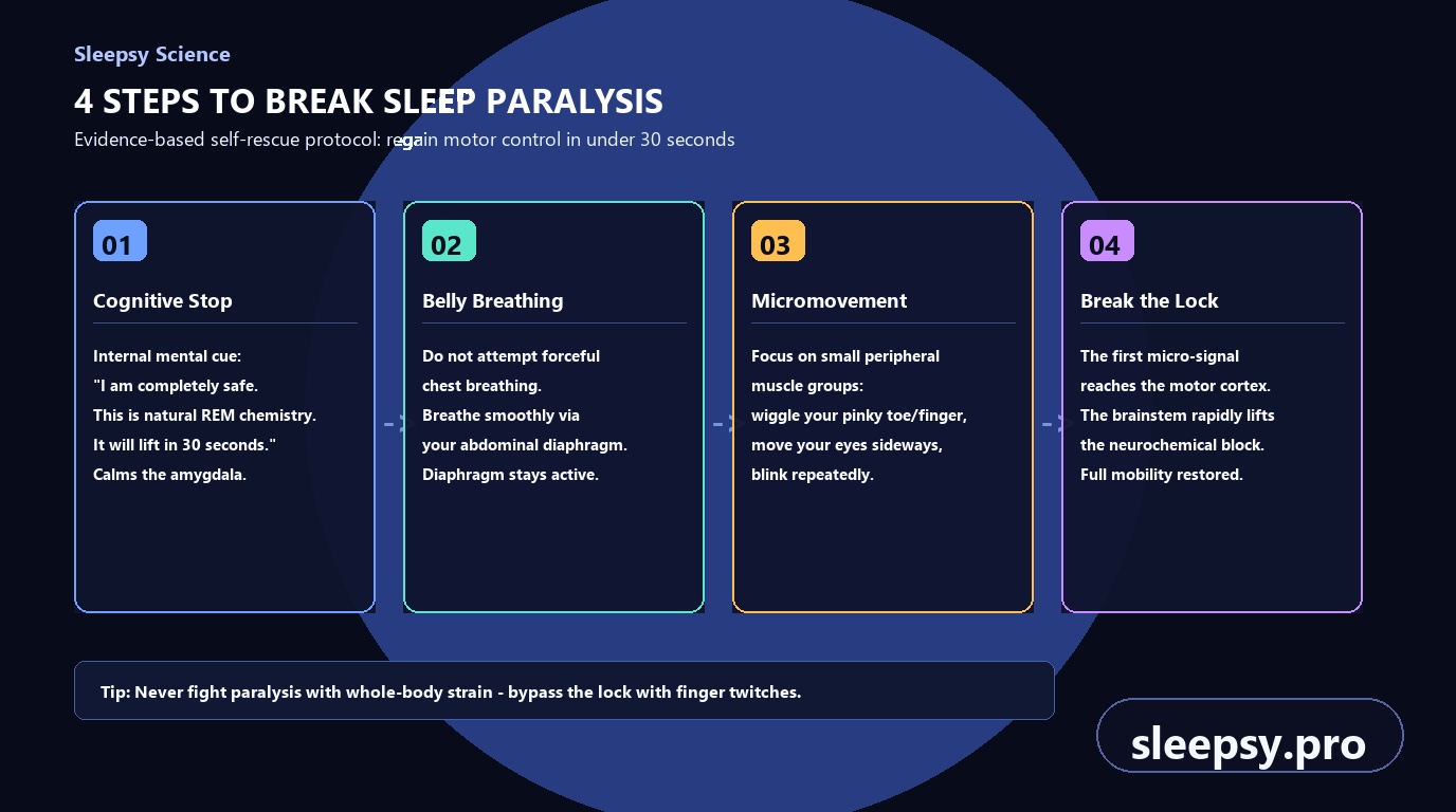
Follow this structured, 4-step emergency protocol whenever an episode begins:
- Cognitive Stop Cue. Immediately tell yourself: “My body is completely safe. This is normal REM chemistry, GABA and glycine. It will lift within 30 seconds.” This conscious rationalization arrests amygdala panic.
- Smooth Diaphragmatic Breathing. Abandon attempts to take forced, deep chest breaths. Allow your diaphragm to handle regular nasal breathing. The autonomic respiratory center performs flawlessly without conscious effort.
- Target Peripheral Micromovements. REM atonia incapacitates large proximal muscles, but largely spares cranial oculomotor pathways and distal digits. Rapidly blink your eyelids, roll your eyes from side to side, or channel all your attention into twitching the tip of a single pinky finger.
- Release the Lock. The moment that small distal muscle twitches, a clear proprioceptive signal travels back up to the somatosensory cortex. The brainstem detects the movement, lifts the biochemical gate, and restores voluntary control across the entire body within seconds.
Key Takeaways
- Sleep paralysis is a brief, benign asynchrony between a conscious brain and resting skeletal muscles.
- REM atonia is mediated by inhibitory neurotransmitters GABA and glycine, designed to shield you from acting out dreams.
- Apparitions and chest tightness stem from an adrenaline surge in the amygdala combined with natural diaphragmatic breathing.
- Supine posture, sleep deprivation, and chronic psychological stress are the primary behavioral triggers.
- You can break an episode in under half a minute through slow abdominal breaths and focused micromovements of a single finger or eye blinking.
Sleep paralysis is neither a medical danger nor an occult mystery—it is simply a momentary delay in lifting nature’s protective motor fuse while your waking mind greets the day.
If fragmented rest, abrupt nocturnal awakenings, or morning grogginess continue to disrupt your daily focus, targeted sleep tracking can pinpoint the underlying patterns. The Sleepsy intelligent sleep analysis platform helps you map circadian phases, neutralize nocturnal stress triggers, and restore restorative, undisturbed sleep every night.